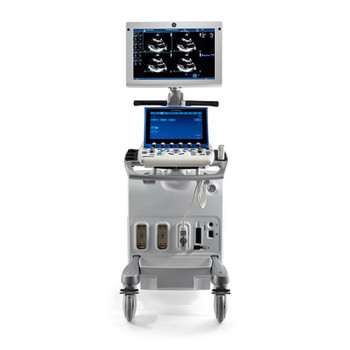
Difference Between Vivid S70 And Vivid S70n
March 21, 2019The GE Vivid line of ultrasound equipment is GE’s Cardiovascular/Cardiac/Cardiology line of ultrasound systems. The Vivid systems can help you provide ultrasound services across a wide variety of cardiology applications, like Transesophageal, Echo, Color Doppler, and many more.
Acura 3.2TL vs. Chrysler 300M, Lexus ES300, Mazda Millenia S, and Five More Entry-Luxury Sedans Ennui and Upward!: A fitful search in the New York woods for passion and personality at a price of. The major difference between the two systems is that the GE Vivid E90 does not have 4D echocardiography. However, the newly added dedicated beamforming software of the GE Vivid E90 provides potentially the best 2D and Doppler cardiac image quality in the market, which an operator can expect by combining cSound and the M5Sc XDclear transducer.
From lightweight, compact and portable to next-generation 4D technology that can be incorporated into your daily routine, GE offers a variety of products to fit a wide range of cardiovascular needs and applications at every budget level. Read below to learn the differences between the GE Vivid cardiovascular ultrasound systems and learn which one is best for you! Part 2 will cover the mid-range Vivid family ultrasound systems. GE Vivid Mid-Range SystemsGE’s mid-range Vivid ultrasound systems are console-based systems with high-powered performance. These cardiac ultrasound systems are targeted at cardiologists and other physicians with smaller practices who need a small-footprint system with big-footprint power. In this line of systems, you have the Vivid Signature-Series and the Vivid T-Series.GE Vivid S5The cardiovascular ultrasound systems combine miniaturization and versatile diagnostic solutions with impressive performance and excellent image quality.

Vivid S70n V202
It’s a console-based system with a portable, sleek design. Advanced functions, an innovative ergonomic design, and broad shared-service capabilities—including vascular, abdominal, pediatric/fetal, and OB—allowing you to keep the focus on your patients. GE Vivid S6The is the higher-end system in the Signature-Series line. Between the Vivid S5 and, the only major difference is the Vivid S6 can run TEE (Transesophageal) ultrasound probes and the S5 cannot. The ergonomic design and easy-to-use features of GE’s Vivid S6 allow you to take workflow and productivity to the next level.
(Read a 2018 update on cardiac ultrasound technologies )There were some big innovation trends in cardiac ultrasound in 2015. These included the launch of the first premium all touchscreen ultrasound system; the introduction of artificial intelligence to speed, simplify and make cardiac echo more reproducible; and a big advancement in image quality, approaching that of computed tomography (CT) scans.Additionally, vendors continue to improve overall image quality, ergonomics and workflow and automation on premium-tier systems to handheld, point-of-care (POC) solutions. Vendors also are concentrating efforts to deliver more affordable systems due to the current economic and healthcare reform climate.
“Hospitals can’t always buy the premium system, nor does everyone need a premium system,” said Jon Brubaker, MBA, RCVT, ultrasound technology analyst for MD Buyline. “Customers that are doing well have a broad product line, and vendors can go to them and say ‘You may want to mix this product with this product.’ ”There has been a slow uptake in 3-D ultrasound in recent years, partly due to high expense of these systems and because of the large amount of volumetric image data that first need to be processed into the required 2-D views. Prior to recent automation software, this was very time consuming. “People are saying, ‘I’m perfectly happy with 2-D ultrasound, why should I move to that? It takes more time and I have to learn how to do it,’” Brubaker explained.


“But I think we are reaching that stage where it is going to be the standard of care.” He believes what will ultimately push 3-D echo technology past that tipping point is its ability to reduce or eliminate operator variability in scanning and interpretation. Similar to a CT scan, 3-D ultrasound captures a cone shaped volume of image data, which can be sliced and viewed on any plane. This means echo is no longer dependent on the sonographer to get the best possible viewing plane. “It’s one of the most significant things you have to overcome.